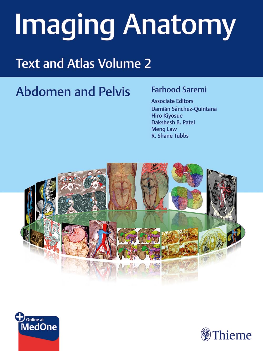
Imaging Anatomy Text and Atlas Volume 2: Abdomen and Pelvis 1st Ed.
Imaging Anatomy Text and Atlas Volume 2: Abdomen and Pelvis 1st Ed.
Couldn't load pickup availability
Normal imaging anatomy and variants, including diagnostic and surgical anatomy, are the cornerstones of radiologic knowledge. Imaging Anatomy: Text and Atlas Volume 2, Abdomen and Pelvis is the second in a series of four richly illustrated radiologic references edited by distinguished radiologist Farhood Saremi. The atlas is coedited by esteemed colleagues Damián Sánchez-Quintana, Hiro Kiyosue, Dakshesh B. Patel, Meng Law, and R. Shane Tubbs with contributions from an impressive cadre of international authors. Succinctly written text and superb images provide readers with a virtual, user-friendly dissection experience.
This exquisitely crafted atlas combines fundamental core anatomy principles with modern imaging and postprocessing methods to increase understanding of intricate anatomical features. Twenty-two concise chapters cover the abdominal wall, alimentary tract, liver, biliary system, pancreas, spleen, peritoneum, genitourinary system, pelvic floor, neurovasculature, and surface anatomy.
Share

